
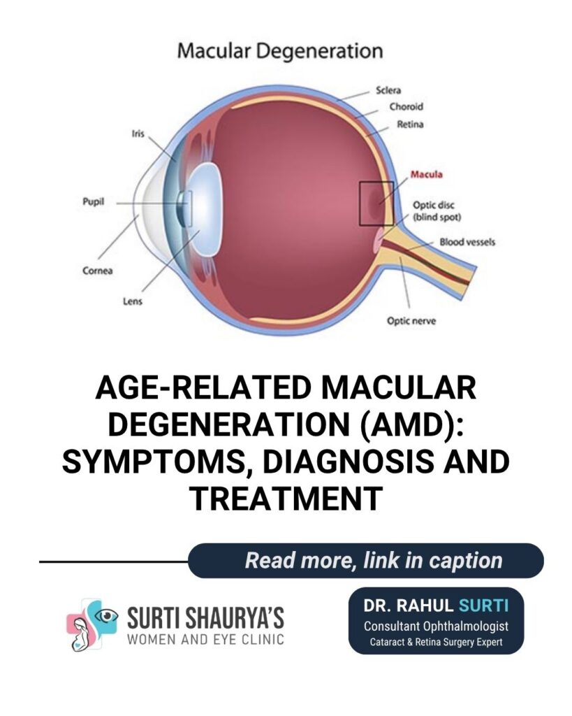
Age-related macular degeneration (AMD) is a common eye condition that affects the macula—the tiny, highly sensitive area at the center of the retina. The macula is responsible for crisp, straight-ahead vision, enabling essential daily activities like reading, recognizing faces, driving, watching TV, and using a mobile phone.
While AMD primarily compromises central vision, it generally spares peripheral (side) vision, meaning it rarely leads to complete darkness or total blindness. However, advanced stages can severely impact quality of life and independent living.
AMD develops due to a combination of aging, genetic predisposition, and environmental factors. As the eye ages, tiny cellular waste deposits called drusen can accumulate underneath the retina, leading to degeneration of the retinal pigment epithelium (RPE) and photoreceptor cells.
Key risk factors include:
- Age: Risk increases significantly after age 50 to 60.
- Family History: Genes play a strong role; having a close relative with AMD increases your risk.
- Smoking: The single most critical modifiable risk factor. Smoking doubles the risk of developing AMD and accelerates progression.
- Cardiovascular Health: High blood pressure, elevated cholesterol, and vascular disease contribute to retinal tissue stress.
- Diet & Lifestyle: Poor nutrient intake, lack of exercise, and obesity increase susceptibility.
AMD presents in two primary forms, which behave differently and require distinct approaches to monitoring and clinical care.
Feature | Dry AMD (Non-Neovascular) | Wet AMD (Neovascular) |
Prevalence | Accounts for ~80–90% of all cases | Accounts for ~10–20% of cases |
Mechanism | Gradual thinning of the macula and drusen buildup | Growth of abnormal, fragile blood vessels that leak fluid or blood |
Progression Speed | Slow, gradual progression over years | Rapid, sudden changes in central vision |
Key Warning Sign | Mild blurring, need for brighter light when reading | Straight lines appearing wavy or distorted (metamorphopsia) |
Primary Management | Lifestyle shifts, monitoring, AREDS2 supplements | Prompt intravitreal anti-VEGF injections |
Stages of Dry AMD
- Early: Drusen are small or few. Often produces no noticeable visual symptoms.
- Intermediate: Drusen become larger. You may notice mild blurring or require brighter light for fine tasks.
- Advanced: Significant loss of retinal cells and areas of tissue breakdown (geographic atrophy), causing persistent dark patches in central vision.
In its earliest phases, AMD may be completely asymptomatic. As tissue alterations progress, you may experience:
- Blurring or fading of central vision
- Difficulty reading, recognizing faces, or seeing fine details
- Reduced visual contrast or reduced vividness of colors
- Distortion (Metamorphopsia): Straight edges—such as door frames, window blinds, or line spacing on paper—look bent, wavy, or crooked
- A growing blurry or dark spot (scotoma) right in the middle of your field of view
- Difficulty adapting to dim or low-light conditions
Note: AMD can affect one eye much more severely than the other. Because the healthy eye compensates, early vision loss often goes unnoticed until you cover one eye at a time to test each individually.
A comprehensive dilated eye exam is essential for detecting AMD early. Specialist diagnostic tools include:
- Visual Acuity Testing: Evaluates distance and near clarity using standardized letter charts.
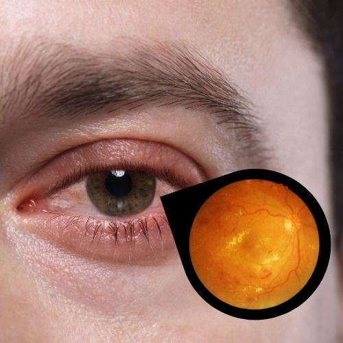
- Dilated Fundus Examination: Allows the specialist to directly view the macula for drusen, fluid, atrophy, or hemorrhage.
- Optical Coherence Tomography (OCT): High-resolution, cross-sectional imaging that reveals microscopic layers of the retina, fluid accumulation, swelling, and structural changes.
- Amsler Grid Test: A simple grid pattern used to detect subtle visual distortions or blind spots.
- Fluorescein Angiography: Involves injecting a harmless fluorescent dye into a vein in the arm to map blood flow and highlight leaking, abnormal blood vessels beneath the retina.
While current treatments cannot completely reverse existing permanent retinal damage, modern therapies can slow progression and protect remaining vision.
Managing Dry AMD
- Regular Monitoring: Scheduled tracking with OCT scans to watch for progression to wet AMD.
- Lifestyle Interventions: Smoking cessation, balanced nutrition rich in antioxidants (leafy greens, omega-3 fatty acids), and blood pressure control.
- AREDS2 Supplements: Specific high-dose combinations of vitamins C and E, zinc, copper, lutein, and zeaxanthin are clinically proven to slow progression in patients with intermediate or advanced single-eye AMD.
Treating Wet AMD
- Anti-VEGF Injections: Medications targeting Vascular Endothelial Growth Factor (a protein that stimulates abnormal vessel growth) are injected directly into the vitreous cavity of the eye. These medications shrink abnormal vessels, clear fluid, and stabilize vision.
- Treatment Frequency: Initial monthly injections are often followed by tailored “treat-and-extend” regimens based on visual acuity and OCT imaging results.
- Photodynamic Therapy (PDT): A specialized laser combined with a light-activated intravenous drug used in select complex cases.
Is AMD the same as a cataract?
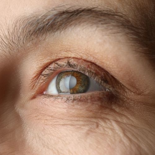
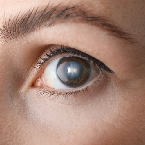
No. Cataracts cause cloudiness in the natural lens at the front of the eye and can be cured with routine surgery. AMD affects the light-sensitive retina at the back of the eye and is a chronic condition. It is common to have both simultaneously.
Can AMD cause total blindness?
AMD affects central vision, leaving peripheral (side) vision intact. While it causes central blind spots, it rarely results in complete darkness.
When should I seek emergency evaluation?
Seek immediate attention from an eye specialist if you experience sudden onset of wavy lines, unexpected central distortion, or a new dark patch appearing in your field of vision.
For patients seeking retinal care in Sion, Mumbai, comprehensive diagnostic evaluations and individualized management plans for AMD are available under the guidance of ophthalmologist Dr. Rahul Surti.
MBBS, MS (Ophthalmology)
FVRS (Fellowship in Vitreo Retinal Surgery)
Advanced Eye Surgeon for Cataract & Retina
Dr. Rahul K. Surti is an experienced eye specialist with expertise in cataract and retina surgeries. He focuses on advanced diagnostic and surgical care to help patients restore clear, healthy vision and
improve their quality of life.
Welcome to Surti Shaurya’s Women & Eye Clinic, a trusted destination in Sion West, Mumbai, for specialized cataract, retina issues, advanced eye surgeries and gynecology, pregnancy care, infertility treatments. Led by Dr Rahul Surti and Dr. Ami R. Surti, the clinic offers compassionate, expert-led care for people of Mumbai.
Contact Details:
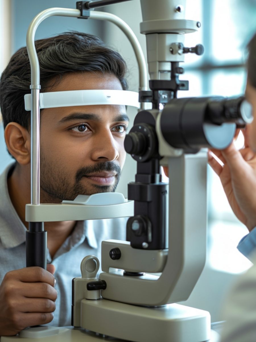
Dr. Rahul Surti offers comprehensive eye care under one roof — from routine check-ups to advanced eye surgeries — ensuring clear, healthy vision for every age.
- Eye Check-Up: Complete vision assessment, refraction test, eye pressure check, and retina evaluation for early detection of eye problems.
- Cataract Treatment: Stitch-less, microincision cataract surgery with advanced foldable and premium IOL options for faster recovery and sharper vision.
- Retina Services: Expert diagnosis and treatment for diabetic retinopathy, macular degeneration, and retinal detachment using advanced laser and imaging technology.
- Advanced Eye Surgeries: Glaucoma, cornea, squint, oculoplasty, and LASIK surgeries performed with precision and the latest microsurgical techniques.
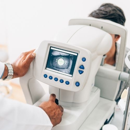
- Computerized Eye Check-Up
- Phaco-emulsification Cataract Surgery
- Sutureless Cataract Surgery
- Refraction
- Contact Lens Clinic
- Squint Evaluation
- Diabetic Retinopathy Check-up
- Retinal Evaluation For Myopia / Retinal Detachment
- Laser Surgeries
- Endoscopic: Sac Surgeries (no Stitch DCR) Lazer Sac Surgery
- Oculoplastry